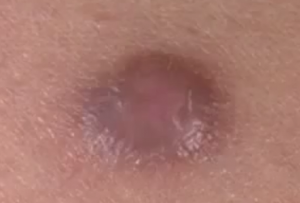
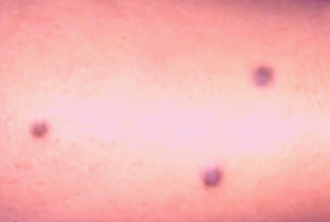
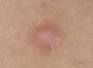
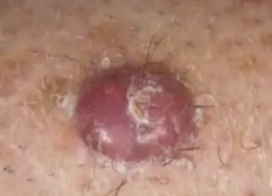
Dermatofibroma is a non cancerous skin growth that typically occurs on the legs. They are small in size and generally grow up to less than half an inch or 1 cm in diameter. Dermatofibroma is mainly made up of fibrous tissue. The condition is also known as sclerosing hemangioma.
The skin growth is a type of urticaria, which is a condition characterized by the development of skin welts along the areas where the affected person has been scratched or stroked. Dermatofibroma generally occurs as a solitary growth but can also occur in multiples.
A dermatofibroma is a circular, brown to purplish growth that can be normally found on the arms and legs. They feel like hard bumps on the skin and are composed of scar tissue. A dermatofibroma contains an abundance of scar-like tissue insider the deeper skin layers, i.e. the dermis.
A dermatofibroma is also occasionally referred to as a fibrous histiocytoma, because of the presence of a benign development of dermal dendritic histiocyte cells
Symptoms of dermatofibroma
Some of the signs and symptoms of dermatofibroma are as follows:
- They normally occur on the lower sections of the legs, but can also be found on the trunk or the arms
- A dermatofibroma can vary in size and can range from being as small as a BB pellet to growing as big as a fingernail.
- It is elevated from the skin and tends to bleed when injured
- One may observe pinkish, reddish, purple brownish or gray patches occurring in the affected parts of skin
- When pinched, they tend to dimple inwards
- The color may alter with the passage of time
- People with darker skin may have darker dermatofibromas
- They may sometimes be sensitive and become itchy on touching
- A dermatofibroma usually does not cause any pain, but can become itchy, tender and painful
- The condition can appear at any age, but tends to affect women more frequently as compared to men. Dermatofibromas normally tend to occur as a single growth, but occasionally can appear as many.
It is important to note that even though dermatofibromas are harmless, one must consult a doctor for correct diagnosis of any new growth on the skin, particularly one which is black or dark brown, or changes shape, size or color. In case, a growth grows rapidly, or bleeds, or results in pain, then immediate medical attention is necessary.
Causes of dermatofibroma
- The precise cause of dermatofibroma is still a matter of research. The condition first grows as a solid papule or a button-like nodule. Such lesions may be somewhat tender and their size may change slightly with time.
Some of the risk factors that may increase the vulnerability to developing a dermatofibroma are listed below:
- Individuals with a family history of the condition may be prone to developing the condition
- Any type of minor injury, like a prick from a thorn can also increase the susceptibility to developing a dermatofibroma
- Women are at greater risk to developing the condition than men
- It can also result from an insect bite
- Dermatofibromas rarely occur in children and generally tend to affect middle aged adults
Dermatofibroma removal and treatment
- Treatment is rarely needed for dermatofibroma. Some individuals affected by the condition may choose to remove the dermatofibromas for cosmetic reasons or when it occurs at an inconvenient place. For example, dermatofibromas that are prone to getting nicked while shaving or which get irritated by apparels. Also, itchy and painful dermatofibromas may require treatment.
- In case of an indefinite diagnosis of the condition, doctors may recommend removal of a piece for tissue analysis.
- A dermatofibroma can be easily removed via surgery. However, since the growths tend to have deeper roots, their excision occurs below the top layers of the skin. Hence, such a process normally results in scarring.
- Alternatively, people affected by dermatofibroma can also opt for different treatment procedures to get rid of the growth. Freezing therapy involves the use of liquid nitrogen to freeze off the top end of a dermatofibroma and thus destroy it. Additionally, the top half of dermatofibromas can also be scraped off with the help of a surgical knife.
- It may be noted that the above procedures may not leave a scar, but since the processes involve removal of only the top half of a dermatofibroma with the deeper layers remaining intact, the chances of the condition reoccurring is elevated. In such cases, affected people have to go for another session of dermatofibroma removal, when it recurs.
- In rare cases, a particular type of skin cancer that initially looks like a dermatofibroma can travel to other body regions. This form of skin cancer is known as DFSP or dermatofibrosarcoma protuberans. Hence, all cases of skin growths have to be initially checked by a doctor, for accurate diagnosis.
Dermatofibroma Pictures


Whats up very nice web site!! Man .. Excellent .. Superb .. I will bookmark your web site and take the feeds additionally…I am satisfied to search out so many helpful info right here in the post, we want work out extra techniques in this regard, thank you for sharing. . . . . .