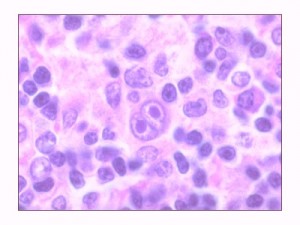
If the doctor suspects the presence of swelling or signs of lymphoma, he/she may ask a volley of questions about the present and past illnesses, consumption of medications if any, family history and the lifestyle of the patient. Afterwards, the health care provider may go for a series of complete medical examinations for further clarification about lymphoma. The doctor may also refer the patient to consult specialists in blood disorders and cancers.

Lymphoma diagnosis includes the following:
Blood tests
Blood samples drawn for various tests such as:
- Evaluating functions of blood cells and vital organs such as kidneys and liver.
- Determining blood chemicals or enzymes. Higher presence of LDH in suspected NHL cases may indicate a hostile form of the disorder.
- More tests may be conducted to know about subtypes.
Biopsy
- From among several methods, the removal of sample tissue from a lump by a pathologist for examination is called as biopsy.
- Small sample of mass is removed by inserting a hollow needle. The procedure is known as core-needle biopsy.
- Core-needle biopsy does not provide a good quality sample. Hence doctors prefer a surgical biopsy, wherein the swollen lump node is removed through a small incision in skin.
- If the mass is deep inside the body, the tissue sample is taken by laparoscopy. The procedure involves making a tiny incision in skin and inserting a light fitted thin tube with camera at the end. Pictures of insides of the body are sent by camera to a video monitor. Tissue can then be withdrawn from body by the help of a small cutting tool fitted on the end of laparoscope.
- Pathologist’s microscope examination report will specify the lymphoma, along with its classification.
Imaging studies
If, inspite of the presence of symptoms, no palpable mass is found, then imaging studies are taken to determine the presence of mass and for guidance to direct a biopsy if required. Some imaging studies include:
- X-rays: Sometimes a simple X-ray can detect lymphoma.
- MRI scan: MRI gives much better three dimensional details than CT scan, especially for brain and the spinal cord.
- CT scan: It gives a three dimensional and detailed view which helps in easy lymphoma diagnosis.
- Gallium scan: Lymphomas tend to accumulate a substance called gallium when it is injected into a body. A scan, after injecting a small quantity of radioactive gallium will show the areas of gallium collection, indicating a tumor.
- Lymphangiogram: It gives the image of lymphatic system by tracing a dye that moves through the system. It is an outdated method and has been replaced by CT scan, MRI or PET.
- Positron-emission tomographic (PET) scan: It is a new choice, alternative to lymphangiogram and gallium scan, for locating areas of body affected by lymphoma. A small quantity of radioactive substance is injected in body, and traced on PET scan.
Bone marrow examination
- To confirm that the bone marrow is not affected by lymphoma, a biopsy of bone marrow is collected.
- Normally samples are taken from the hip bone.
- The bone marrow is examined by pathologist under a microscope.
- Presence of particular types of abnormal B and T lymphocytes in bone marrow will confirm the lymphoma.
Other tests
- Lumbar puncture: A sample of cerebrospinal fluid surrounding the brain and spinal cord is collected by spinal tap method to confirm the presence of lymphoma cells in central nervous system.
- Organ function tests: Tests like echocardiogram or MUGA are taken before treatment to see whether the vital organs can withstand the side effects of therapy.
Staging
Classification of cancer types, size and its stage may help oncologist to determine the type of treatment and the chances of a cure.
Staging of lymphomas depend on the results of imaging studies and the related tests. Non-bulky lymphoma has better chances of cure than bulky ones.
The newest NHL classification system takes into account microscopic picture, location, genetic, molecular features and grade of lymphoma.
- Low grade: It is also called as “indolent” lymphomas because of its slow growth. It does not require urgent treatment, unless it affects functions of organs.
- Intermediate grade: These are aggressively growing lymphomas and need immediate treatment. These are usually curable.
- High grade: These are aggressively growing lymphomas. Need immediate and intensive care and their chances of curability are lesser.
The “staging” and evaluation of lymphomas are similar for HL and NHL.
- Stage 1 (early lymphoma): Lymphoma is located in single lymph node region.
- Stage 2 (locally advanced lymphoma): Lymphoma is located in more lymph node regions, all on the same side of diaphragm.
- Stage 3 (advanced lymphoma): Lymphoma affecting two or more lymph node regions
- Stage 4 (widespread disease): Lymphoma is wide spread outside lymph nodes and spleen covering other areas and systems.
Classification of HL and NHL with letters
- Patients who do not suffer from fever and weight loss are classified as “A”, while those who experience them are classified as “B”.
- If the tumor spreads directly from a lymph node into an organ outside the lymphatic system, then it is classified as “E”. If the spleen is also involved, then it is classified as “S”
Exceptional post but I was wanting to know if you could write a litte more on this topic? I’d be very grateful if you could elaborate a little bit further. Appreciate it!